Equipment
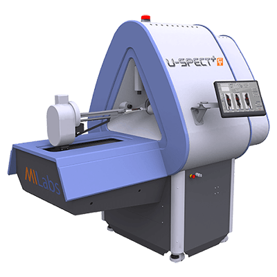
- MILabs U-SPECT+/CT Single Photon Emission Computed Tomography (SPECT) with x-ray Computed Tomography system

Example Applications
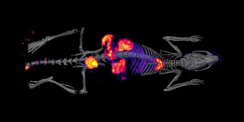
- Study drug toxicity longitudinally in vivo after registering SPECT images with MRI anatomical maps
- Assess kidney health in transplant models
- Use NIS reporter gene for cell tracking experiments
Recent SPECT publications
Targeted design of a recombinant tracer for SPECT renal imaging
Liu P, .. Jin J. Theranostics. 2021;11(18):9118-32. Continue Reading Targeted design of a recombinant tracer for SPECT renal imaging
Whole-body imaging of cell death provides a systemic, minimally invasive, dynamic and near-real time indicator for chemotherapeutic drug toxicity.
Johnson SE, … Zhao MW. Clin Cancer Res 2019 Feb 15;25(4):1331-1342. DOI link Continue Reading Whole-body imaging of cell death provides a systemic, minimally invasive, dynamic and near-real time indicator for chemotherapeutic drug toxicity.
Technical Details
SPECT/CT
A state-of-the-art micro MILabs U-SPECT+/CT Single Photon Emission Computed Tomography (SPECT) with x-ray Computed Tomography was installed in CAMI in August 2014. It has higher spatial resolution than its competitors (0.25 mm) and higher sensitivity (as high as 12000 cps/MBq), using three NaI(TI) 472 × 595 detector panels. Three general purpose collimators are available; one each for mice, rats, and rabbits. There are two extra high resolution collimators, one each for mice and rats. The CT system has 12-bit, 1280 × 1024 detector, capable of 80 µm voxel size. An animal monitoring system allows for cardiac and respiratory gated image acquisition (M2M BioVet). Image acquisition is performed on a quad-core CPU computer and image reconstruction on a 32 core workstation.

