Equipment
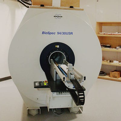
- 9.4T Bruker Biospec MRI System: for hardware-intensive experiments such as spectroscopy, diffusion imaging and X-nucleus experiments;with Autopac laser positioning system enabling reproducible subject positioning for timecourse studies
- 7T Bruker Pharmascan MRI System: for high-throughput basic imaging experiments
Techniques
- High resolution anatomomical imaging
- T1 and T2 relaxometry
- Diffusion imaging (DWI, DTI)
- Dynamic contrast enhanced imaging (DCE)
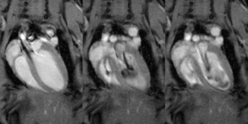
- Cardiac functional imaging
- Multimodality imaging
Example Applications
- Track longitudinal changes in tumor microenvironment in response to drug treatment
- Measure changes in cardiac function (cardiac output, left ventricular mass, wall motion, etc.) after a myocardial infarction
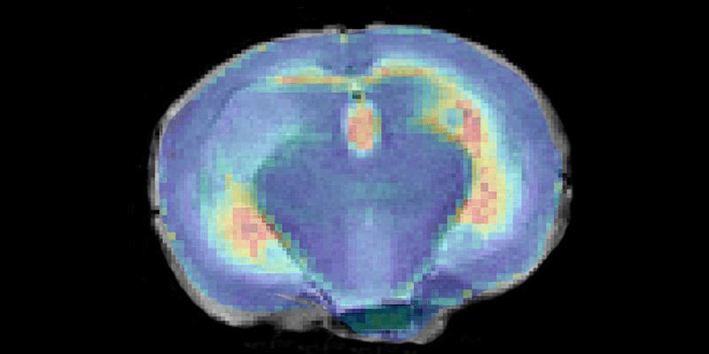
- Track labeled tumor cells in the brain
- Create a map of abdominal organs that can be registered with SPECT images for organ-level quantification of SPECT signal
- Quantify and visualize changes in body fat distribution in response to experimental manipulation
- Characterize the performance and biodistribution of novel MRI contrast agents at high field
Recent MRI publications
The Therapeutic and Diagnostic Potential of Amyloid β Oligomers Selective Antibodies to Treat Alzheimer’s Disease
Viola KL, .. Klein WL. Frontiers in Neuroscience. 2022;15(1798). Continue Reading The Therapeutic and Diagnostic Potential of Amyloid β Oligomers Selective Antibodies to Treat Alzheimer’s Disease
Wireless, Battery-Free Subdermally Implantable Photometry Systems for Chronic Recording of Neural Dynamics
Burton A, … Gutruf P. Proc Natl Acad Sci U S A. 2020 Feb 11;117(6):2835-2845. Continue Reading Wireless, Battery-Free Subdermally Implantable Photometry Systems for Chronic Recording of Neural Dynamics
Self-Immolative Activation of β-Galactosidase-Responsive Probes for In Vivo MR Imaging in Mouse Models.
Lilley LM, … Meade TJ. Angew Chem Int Ed Engl. 2020 Jan 2;59(1):388-394. DOI link Continue Reading Self-Immolative Activation of β-Galactosidase-Responsive Probes for In Vivo MR Imaging in Mouse Models.
Technical Details
9.4T MRI
The 9.4T Bruker Biospec MRI system (30 cm bore) is designed for hardware-intensive experiments such as high resolution imaging, functional imaging, and diffusion imaging. The large gradient set has an internal diameter of 20 cm and a gradient strength of 200 mT/m, and is suitable for imaging larger rodents such as rabbits and guinea pigs. The gradient insert with 12 cm diameter and a gradient strength of 450 mT/m is suitable for imaging mice and rats. An AutoPac laser positioning system enables rapid and precise sample positioning. The system has four receive channels for parallel imaging, and four transmit channels, including one for X-nuclei.
Available coils:
Transmit/Receive Volume coils:
- 23mm quadrature coil for mouse head
- 25mm quadrature coil for phantoms
- 38mm quadrature coil for rat head and mouse body
- 72mm quadrature coil for rat body
- 72mm 1H/19F dual-tune linear coil
Receive-only Surface coils (used in conjunction with 72mm quadrature volume coil as transmitter)
- 4 channel phased array (2×2) surface coil for mouse brain
- 4 channel phased array (2×2) surface coil for rat brain
- 4 channel phased array (1×4) surface coil for rat heart
7T MRI
The 7T Bruker PharmaScan MRI system (16 cm bore) is designed for high throughput experiments for imaging anatomy and basic function. Its gradient set has a 9 cm bore suitable for imaging mice and rats, and a strength of 120 mT/m. Four receive channels enable parallel imaging.
Available coils:
Transmit/Receive Volume coils (fixed tune/match):
- 23mm quadrature coil for mouse head
- 35mm quadrature coil for mouse body
- 60mm quadrature coil for small rats
Receive-only Surface coils (used in conjunction with 72mm linear transmit-only volume coil)
- 4 channel phased array (2×2) surface coil for mouse brain
- 4 channel phased array (2×2) surface coil for rat brain


