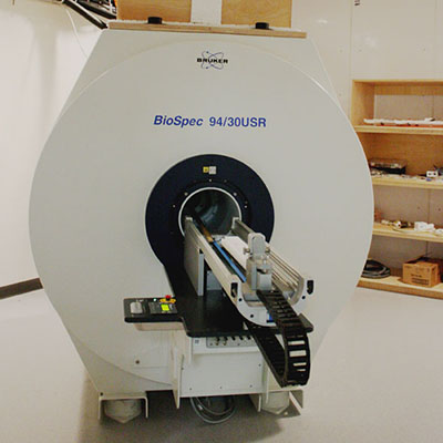
Bruker Biospec 9.4T MRI
The 9.4T Bruker Biospec MRI system is designed for hardware-intensive experiments. It is equipped with an Autopac laser positioning system enabling reproducible subject positioning for timecourse studies.
Example applications include high resolution anatomic imaging in mouse and rat body, diffusion imaging (DWI, DTI) of brain or tumors, dynamic contrast enhanced imaging (DCE) of tumors, cardiac functional imaging, and multimodality imaging with other CAMI instruments.